Here is the video PBS recently made about Parkinson’s disease called My Father, My Brother, and Me. From what I’ve watched so far, it’s done a good job putting a face to Parkinson’s disease while also focusing on the research and clinical aspects of it.
Hippocampus Anatomy Video
To follow up my previous post on the hippocampus, here’s a video posted by drbobrd on YouTube. He uses a model of a brain to explain some brain anatomy, including the hippocampus and fornix.
The Hippocampus in 400 Words
Within the temporal lobe of the brain is an elongated structure called the hippocampus. Some people have compared its shape to that of a seahorse (the word hippocampus comes from the Greek {hippos + campos}, which roughly means “seahorse”). This structure is special for a number of reasons. One is its role in memory encoding and consolidation.

From cytoarchitectonic standpoint, the hippocampus is special because unlike the surrounding cortex, it consists of only three layers instead of six. The hippocampus is phylogenetically an old part of the cortex, which means that it is an older branch on the evolutionary tree, whereas the rest of the cortex (more accurately called the neocortex), especially cortex of the frontal lobes, is a much newer development.
The hippocampus resides within the medial portion of the temporal lobe. It is continuous with the parahippocampal cortex, entorhinal cortex (the hippocampus receives its main input from this cortex), and perirhinal cortex.
The hippocampus sends white matter tracts off its dorsal and posterior portions (the hippocampus also communicates through other tracts and pathways – this circuit is not the only output of the hippocampus). These white matter tracts are the fimbria of the hippocampus (technically, the fimbria are the “offshoots” of the alveus of the hippocampus). The fimbria proceeds upwards from the posterior portion of the hippocampus, at which point it ceases to be the fimbria and is called the fornix.
The fornices (plural of fornix) are prominent white matter tracts passing above the thalamus and medially in the brain. The fibers travel forward, then turn downward just posterior to the anterior commissure (a white matter tract that connects both hemispheres) to terminate in the mammillary bodies, two bumps on the ventral side of the brain. They are part of the hypothalamus of the brain. From there, the pathway courses upward through the mammilothalamic tract (MTT) to the anterior nucleus of the thalamus. From there axons course to the cingulate gyrus, then to the underlying cingulum (large white matter tract), and back to the hippocampus (via the parahippocampal and entorhinal cortices). This circuit is part of the limbic system and is called the Papez circuit. This circuit is important for emotion and memory.
An Introduction to and Overview of the Brain

The human brain is a wondrous thing. It is the single most complex organ on the planet. It sits atop the spinal cord. Gazing upon the brain, one sees four main distinct areas – two roughly symmetrical hemispheres, a cerebellum stuck up underneath the posterior part of the brain, and a brainstem sticking out and down from the middle of the brain. Each cerebral hemisphere is divided into four visible lobes: frontal, temporal, parietal, and occipital. The frontal lobes jut out at nearly a 90 degree angle from the spinal cord and are the largest part of the human brain. The temporal lobes stick out the sides of the brain, like thumbs pointing forward at the side of a fist. The parietal lobes are harder to distinguish. They are just posterior to the frontal lobes and dorsal to (above) the temporal lobes. The occipital lobes are at the very back of the brain, like a caboose on a train.
The outside of the brain is covered with a series of bumps and grooves. The bumps are called gyri (sing. gyrus) whereas the grooves are called sulci (sing. sulcus). This outside part of the brain is filled with tiny cell bodies of neurons, the main functional cell of the brain. Some people estimate that there are 100 billion neurons in the central nervous system (brain + spinal cord). This outer layer of the brain is called the cortex (which means “bark”). The cortex is only about 5mm thick, or about the thickness of a stack of 50 sheets of copy paper, yet it is responsible for much of the processing of information in the brain.
At room temperature the brain is the consistency of warm cream cheese. If removed from the skull and placed on a table, it would flatten and widen out a bit, like jello that is warming up. The brain is encased in a series of protective sheaths called meninges. The outermost encasing is called the dura mater (L. “tough mother”), which is thick and tough and is attached to the skull. The next layer in is softer. It is called the arachnoid layer; it adheres to the brain. Just underneath this layer is where cerebrospinal fluid (CSF) flows. This fluid is produced in holes in the middle of the brain called ventricles. CSF helps cushion the brain as well as remove waste products from the brain. Underneath this is a very thin and fine layer called the pia mater (L. “soft mother”), which adheres directly to the cortex and is difficult or impossible to remove without damaging the cortex. These three layers of meninges serve to protect the brain.
The brain can be roughly split into three functional areas, each one more “advanced” than the previous. The brainstem (and midbrain), which includes such structures as the medulla, pons, and thalamus, activates and regulates the general arousal of the cortex. Damage to the brainstem often results in coma or death. The next rough functional area is the posterior portion of the brain (parietal and occipital lobes and portions of the temporal lobes). This area is heavily involved in sensory processing – touch, vision, hearing. It sends information to other parts of the brain largely through the midbrain structures. The last functional area includes the frontal lobes. This area can regulate all other parts of the brain but is essential for goal-setting, behavior inhibition, motor movements, and language. The frontal lobes are the most advanced area of the brain and arguably the most important for human functioning – for what makes us human. In summary the three areas roughly are responsible for:
- Overall arousal and regulation
- Sensory input
- Output, control, and planning
Underneath the cortex is a large area of the brain that looks white. This area is comprised of the axons of the neurons of the cortex and subcortical structures. These axons are the pathways between neurons – like superhighways connecting cities. The axons look white because the majority are covered with a fatty tissue called myelin. Myelin helps axons work more efficiently and transmit more quickly. The white matter of the brain is as important for normal brain functioning as the gray (neurons) matter is.
The brain is energy-hungry. It cannot store energy so it needs a constant supply of nutrients from blood. However, blood can be toxic* to neurons so the brain has to protect itself from the blood and other toxic materials through what is called the blood-brain barrier. This barrier keeps blood cells out of the brain but allows molecules of nutrients (e.g., glucose) to pass into or feed the cells. The entire surface of the brain is covered with blood vessels, with many smaller vessels penetrating deep into the brain to feed the subcortical structures. Deoxygenated blood must be removed from the brain. Veins take the blood out of the brain and drain into venous sinuses, which are part of the dura matter.
The brain works as a whole to help us sense, perceive, interact with, and understand our world around us. It is beautiful in its form and function.
*”Today, we accept the view that the BBB limits the entry of plasma components, red blood cells, and leukocytes into the brain. If they cross the BBB due to an ischemic injury, intracerebral hemorrhage, trauma, neurodegenerative process, inflammation, or vascular disorder, this typically generates neurotoxic products that can compromise synaptic and neuronal functions (Zlokovic, 2005, Hawkins and Davis, 2005 and Abbott et al., 2006).” From Zlokovic, B. V. (2008). The blood-brain barrier in health and chronic neurodegenerative disorders. Neuron, 57(2), 178-201.
Image: Bi Sang by Seung Ji Baek
Brodmann’s Map of the Cortex
I’ll be writing some basic neuroanatomy posts over the coming months (I started with my previous post about the corticospinal tract). I recently finished an intense neuroanatomy course where I learned how much I love basic neuroanatomy. It’s exciting to look at a brain or brain slices and try to figure out what and where different structures are.
In the early 1900s Korbinian Brodmann studied the cytoarchitecture (organization of the cortical layers of neurons) of human and non-human brains. His work was painstaking and thorough. He created a topographic map of the cortex containing 52 (50 in humans) different areas. In my class we were not required to learn all of Brodmann’s cortical areas but had to learn some of the major ones. Brodmann’s Areas (BA) 3,1, and 2 compose the primary somatosensory area of the brain. BA 4 is the primary motor cortex. BA 5 is somatosensory association cortex just posterior to BA 3,1,2. BA 6 is pre-motor cortex, which connects directly to BA 4. BA 7 is more somatosensory association cortex that lies just posterior to BA 5. BA 8 is the frontal eye fields, which among other things is responsible for initiating horizontal eye saccades (i.e., quick movement to the left or right). BA 17 is the primary visual cortex, a credit card sized area that lies both dorsal and ventral to the calcarine fissure in the occipital lobe. This area processes most of the basic visual information. BAs 18 and 19 are visual association cortices. BA 22 is Wernicke’s Area, which is involved in the comprehension of language and is in the dorsal-posterior temporal lobe on the border between the temporal and parietal lobes. BAs 41 and 42 are the primary auditory cortex, which processes auditory information from the cochlea; this lies on the transverse temporal gyrus in the dorsal part of the temporal lobes (it is hidden from view unless the cortex around the Sylvian Fissure is pulled away). BAs 44 & 45 are Broca’s area, which is involved in the production of language and is in the lateral frontal lobes.

The Corticospinal Tract
The corticospinal tract is a descending motor pathway originating in the Primary Motor Cortex (Brodmann’s area 4) and terminating at various levels in the ventral horn of the spinal cord. The corticospinal tract descends through the posterior limb of the internal capsule then down through the cerebral peduncles into the brainstem. In the brainstem the corticospinal tract remains in the ventral portion, passing through the pyramids on its way down. In the caudal brainstem (just above where the spinal cord starts) 90% of the the corticospinal tract decussates (crosses) to the contralateral (opposite) side and continues down through the dorsolateral spinal cord. This portion controls limb movements. The remaining 10% remains in the ventral spinal cord and is largely responsible for bilateral axial (trunk) movement. From the dorsolateral spinal cord, the axon (that started in the cortex) enters the ventral horn of the spinal cord at the appropriate level (e.g., cervical for arms or lumbar for legs) then exits through the ventral root to terminate on the appropriate muscles.

Through this tract, the cortex controls much of the movement of the body; as such, it’s vitally important for our functioning. Damage to the tract results in an upper motor neuron disorder, with paresis (weakness instead of complete paralysis) and the Babinski reflex fairly common symptoms. Soon after damage, a patient might have flaccid paralysis though with little to no movement of the affected limb(s). As the body starts to recover slightly, spastic paralysis usually sets in with jerky, often uncontrolled limb movements. The corticospinal tract is one of the largest pathways in the central nervous system; it’s one of the most important for motor functioning as well.
Another anatomy site
I found a nice but very basic anatomy site (i.e., good for kids). It also has more anatomy than just the brain, with skeletal, heart, and digestive tract anatomy in English and Spanish.
Click on the image to visit the site.

Great Skull Anatomy Site
I stumbled across this wonderful anatomy site that focuses on the skull. You can move your mouse over different parts of the skull to highlight their names. You can also mouse over a structure and have the area of the skull highlighted. This is a wonderful study guide if you have to know the parts of the skull.
|

Split-belt Treadmill as Therapy for Brain-injured Patients
CNN has an interesting article about a split-belt treadmill that is being used for stroke survivors and other people with brain injuries.
The treadmill’s two belts can move independently and even in opposite directions. Doctors and researchers are trying to find any underlying intact neural circuitry by providing unique motor challenges to brain injury patients.
The 3D brain
Technology Review has an interesting article about “new” 3D brain imaging software being developed at Thomas Jefferson University Hospital in Philadelphia, PA (I put “new” in quotation marks because there are other similar programs out there; they might not be as polished but some are even open source). Their software fuses MRI, fMRI, and DTI together to create a fairly comprehensive view of the brain: “The fusion of these different images produces a 3-D display that surgeons can manipulate: they can navigate through the images at different orientations, virtually slice the brain in different sections, and zoom in on specific sections.”
The software looks like it is aimed more at neurosurgeons than researchers (i.e., it probably isn’t free like a lot of MRI image processing software). It does produce amazing images (view the images here) and looks like it could be a very useful tool for at least a qualitative approach to brain imaging.
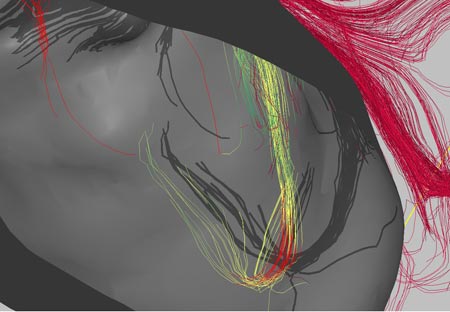
The software is focused a lot on DTI (diffusion tensor imaging) and how the white matter fibers in the brain interact with lesions or tumors. I think that one researcher’s word of caution is important:
“Bruce Fischl, an assistant in neuroscience at Massachusetts General Hospital, says that the idea is ‘interesting’ but cautions that there are a number of levels of ambiguity when talking about connectivity in imaging. ‘Just because you live next to the Mass Pike doesn’t mean that there is an exit,’ he says.”
In other words, don’t get too caught up in the fact that fibers are right by a tumor, they may not really have anything to do with the part of the brain the tumor is most affecting.
In any case, I think that the idea behind this software is amazing. The graphics renderings are impressive (but they are just the pretty pictures – the rendering details may be beneficial in clinical surgery settings but they are not particularly useful in research situations, other than producing nice pictures to go in your publication). This software is very similar to something that I envisioned using a few years ago and I’m glad to see it being developed.
Image credit: Song Lai, Thomas Jefferson University Hospital (borrowed via technologyreview.com)