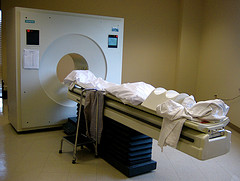
 The positron emission tomography (PET) scan measures blood flow in the brain. This is accomplished by injecting a person or animal with a radioactive isotope (i.e. an unstable atom, usually a variation of oxygen that has a short-half life); this isotope will quickly decay. Founded on the assumption that blood flow will increase in areas of the brain that are in heavy use (such as when a person is viewing an object or reading words or some other cognitive-intensive function), a fair portion of the injected isotopes will end up in the active part of the brain. As the isotopes decay, a positron (a small particle with the exact opposite charge as an electron) is released. This positron will collide with an electron and they will annihilate each other, sending two gamma ray particles in exactly opposite directions. These gamma rays are picked up by the PET scanner, which then determines where they came from in the brain. Since blood should concentrate where the brain is activated, there should be higher levels of isotopes there and this will show up on the scanner in the form of increased levels of gamma rays. The test is usually run twice (once as the control condition and once as the experimental). The difference between the two conditions is measured and any difference should show what area(s) of the brain was (or were) activated.
A PET scan is similar to an fMRI in that both measure blood flow in the brain, which is an indirect measure of brain activity. However, there are advantages and disadvantages to both functional brain imaging methods. PET scans are advantageous in that a person does not have to remain as still as he or she would for the fMRI. Tiny movements can obscure and ruin fMRI data but small movements do not affect PET scans. So, for example, with a PET scan study a researcher could have someone read out loud lists of words but the simple jaw movements would ruin the fMRI data (although this is changing to some degree as image processing becomes more sophisticated; researchers also can modify the task slightly to reduce movement artifacts in fMR images). This is really the main advantage of PET over fMRI.
PET scanning is disadvantaged compared to fMRI because the resolution of the scans is lower. PET scans can measure changes in blood flow in the brain in an area of about 5-10 cubic millimeters. fMRI can resolve down to 3 cubic millimeters and even lower as the machines become more powerful. PET scanning is also much more expensive than fMRI since it takes a special machine, radioactive isotopes, and multiple trials to get a scan. fMRI’s can be done at many hospitals around the world with little or no extra cost because of the prevalence of MRI scanners. Another disadvantage PET’s have are needing radioactive isotopes to work. This isotope can be given only a few times before it is unsafe.
While PET scans were and are better in some situations than fMRI’s, they have many disadvantages overall. With higher cost, lower spatial resolution, and need for isotopes, the disadvantages of PET scans seem to outweigh the advantages.
Image by Muffet.